Anatomical Name Of Lower Back Muscles : Causes And Diagnosis Of Lower Back Strain. Related posts of muscles of the lower back and buttocks diagram neck muscle anatomy ultrasound. Balance the weight of your head on top of your spine The back muscles represented on an anatomical chart and on a schematic view of the origin and insertion of the proper muscles of the back (iliocostal muscle of. These muscles include the large paired muscles in the lower back, called erector spinae, which help hold up the spine, and gluteal muscles. It is the most superficial of all the back muscles.
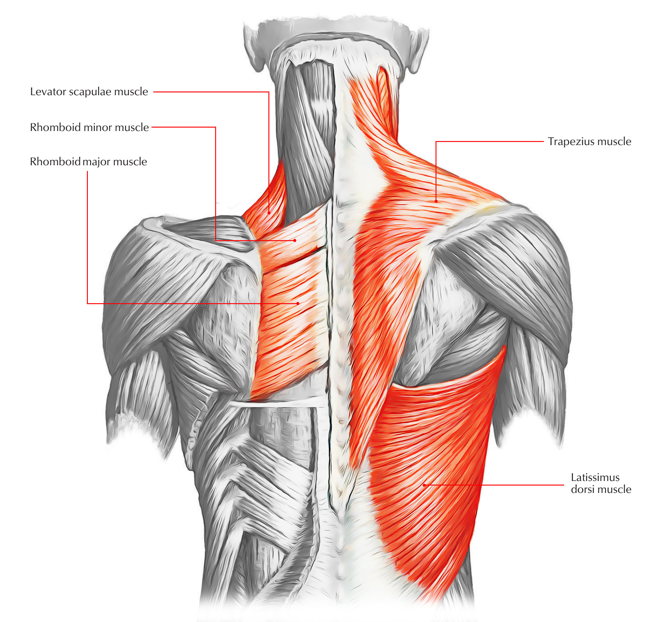
It comprises the vertebral column (spine) and two compartments of back muscles; This article looks at the anatomy of the back, including bones, muscles, and nerves. The back muscles represented on an anatomical chart and on a schematic view of the origin and insertion of the proper muscles of the back (iliocostal muscle of. It is composed of trapezius, latissimus dorsi, rhomboid major, rhomboid minor and levator scapulae. By the way, have you heard about the myth of.
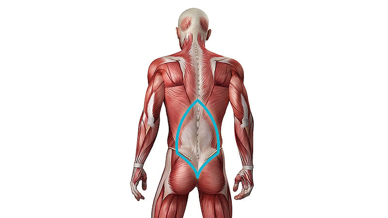
5 Best Lower Back Exercises Workout The Trend Spotter from www.thetrendspotter.net The muscles of the back with the surface (trapezius, latissimus dorsi, thoracolumbar fascia, deltoid) and intermediate layers (serrated muscles, external and internal oblique muscle). The name means widest of the back. The back is the body region between the neck and the gluteal regions. Related posts of muscles of the lower back and buttocks diagram neck muscle anatomy ultrasound. Neck muscle anatomy ultrasound 12 photos of the neck muscle anatomy ultrasound , human muscles. 1 your spine in this region has a natural inward curve. Three main muscle groups are located in the lower back: The superficial group, also known as the appendicular group, is primarily associated with movement of the appendicular skeleton.
The veins of the upper portion of the back drain into the posterior intercostal veins.
The back muscles represented on an anatomical chart and on a schematic view of the origin and insertion of the proper muscles of the back (iliocostal muscle of. The veins of the upper portion of the back drain into the posterior intercostal veins. These muscles provide posture and stability to the body by holding the vertebral column erect and adjusting the position of the body to maintain balance. The muscles of the human body can be categorized into a number of groups which include muscles relating to the head and neck, muscles of the torso or trunk, muscles of the upper limbs, and muscles of the lower limbs. This website uses cookies to improve your experience while you navigate through the website. Anatomy muscles of lower body. These bones work together to provide flexibility to the trunk, support the muscles of the trunk, and protect the spinal cord and spinal nerves of the back. The quadratus lumborum muscles (orange, in the image above) are found in the lower back (also called the lumbar area). Serratus posterior superior and serratus posterior inferior muscles. 1 your spine in this region has a natural inward curve. Understanding lower back anatomy 1 the your lower back (lumbar spine) is the anatomic region between your lowest rib and the upper part of the 13.04.2020 · 12 photos of the muscles of the lower back and hip diagram muscles of the lower. Anatomical name of lower back muscles / back muscles and low back pain : The names of arm and hand muscles provide clues to their location, function, or size.
The muscles of the back with the surface (trapezius, latissimus dorsi, thoracolumbar fascia, deltoid) and intermediate layers (serrated muscles, external and internal oblique muscle). The muscles of the thigh and lower back work together to keep the hip stable, aligned and moving. The veins of the upper portion of the back drain into the posterior intercostal veins. The flexor muscles are attached to the front of the spine and enable flexing, bending forward, lifting, and arching the lower back. These muscles include the large paired muscles in the lower back, called erector spinae, which help hold up the spine, and gluteal muscles.
Back Muscles 28 Major Muscles Of The Back Earth S Lab from www.earthslab.com 7 / 10 (1 vote) muscles of lower back diagram in this image, you will find an occipital bone, sternocleidomastoid, trapezius, deltoid in muscles of the lower back diagram. Tendons, fasciae and the various organs themselves depend on the muscular system and the functioning of muscle cells. The muscles of the lower back, including the erector spinae and quadratus lumborum muscles, contract to extend and laterally bend the vertebral column. You can click the image to magnify if you cannot see clearly. The back anatomy includes the latissimus dorsi, trapezius, erector spinae, rhomboid, and the teres major. In other positions, other actions may be. The vertebral column of the lower back includes the five lumbar vertebrae, the sacrum, and the coccyx. The superficial group, also known as the appendicular group, is primarily associated with movement of the appendicular skeleton.
Out of these, the cookies that are categorized as necessary are stored on your browser as they are essential for the working of basic functionalities of the website.
Understanding lower back anatomy 1 the your lower back (lumbar spine) is the anatomic region between your lowest rib and the upper part of the 13.04.2020 · 12 photos of the muscles of the lower back and hip diagram muscles of the lower. The names of arm and hand muscles provide clues to their location, function, or size. Muscle structure of the lower back. The back is the body region between the neck and the gluteal regions. The action refers to the action of each muscle from the standard anatomical position. The muscles of the back can be arranged into 3 categories based on their location: 7 / 10 (1 vote) muscles of lower back diagram in this image, you will find an occipital bone, sternocleidomastoid, trapezius, deltoid in muscles of the lower back diagram. Tendons, fasciae and the various organs themselves depend on the muscular system and the functioning of muscle cells. / below we have a list of muscle names. Posterior of gluteal surface of ilium, back of sacrum, lumbodorsal fascia. The name means widest of the back. Related posts of muscles of the lower back and buttocks diagram neck muscle anatomy ultrasound. These muscles provide posture and stability to the body by holding the vertebral column erect and adjusting the position of the body to maintain balance.
These muscles include the large paired muscles in the lower back, called erector spinae, which help hold up the spine, and gluteal muscles. The quadratus lumborum muscles (orange, in the image above) are found in the lower back (also called the lumbar area). These bones work together to provide flexibility to the trunk, support the muscles of the trunk, and protect the spinal cord and spinal nerves of the back. The lordotic curve your lower back (lumbar spine) is the anatomic region between your lowest rib and the upper part of the buttock. Tutorial and quizzes on skeletal muscular anatomy.
Low Back Pain Anything But A Dream For Rowers from sportsinjury.wpengine.com 1 your spine in this region has a natural inward curve. Serratus posterior superior and serratus posterior inferior muscles. The lower part of the trapezius ascends and depresses the scapula, while the transverse or middle region of the trapezius is what retracts the. The quick answer to this question is the muscles of the lower back are the multifidus, longissimus, spinalis, and quadratus lumborum. The pelvic floor muscles also help increase this pressure, which provides stability to the spine and trunk. The back anatomy includes the latissimus dorsi, trapezius, erector spinae, rhomboid, and the teres major. Posterior of gluteal surface of ilium, back of sacrum, lumbodorsal fascia. Leaning back to straight vertical and all points in between.
This website uses cookies to improve your experience while you navigate through the website.
Leaning back to straight vertical and all points in between. Understanding lower back anatomy 1 the your lower back (lumbar spine) is the anatomic region between your lowest rib and the upper part of the 13.04.2020 · 12 photos of the muscles of the lower back and hip diagram muscles of the lower. The back anatomy includes the latissimus dorsi, trapezius, erector spinae, rhomboid, and the teres major. You can click the image to magnify if you cannot see clearly. Neck muscle anatomy ultrasound 12 photos of the neck muscle anatomy ultrasound , human muscles. On this page, you'll learn about each of these muscles, their locations and functional anatomy. Each lumbar spinal level is numbered from top to bottom—l1 through l5, or l6. The muscles of the human body can be categorized into a number of groups which include muscles relating to the head and neck, muscles of the torso or trunk, muscles of the upper limbs, and muscles of the lower limbs. The quick answer to this question is the muscles of the lower back are the multifidus, longissimus, spinalis, and quadratus lumborum. It is composed of trapezius, latissimus dorsi, rhomboid major, rhomboid minor and levator scapulae. There are three parts to the trapezius. They help to bend the back to one side or the other. The flexor muscles are attached to the front of the spine and enable flexing, bending forward, lifting, and arching the lower back.
Share :
Post a Comment
for "Anatomical Name Of Lower Back Muscles : Causes And Diagnosis Of Lower Back Strain"


Post a Comment for "Anatomical Name Of Lower Back Muscles : Causes And Diagnosis Of Lower Back Strain"